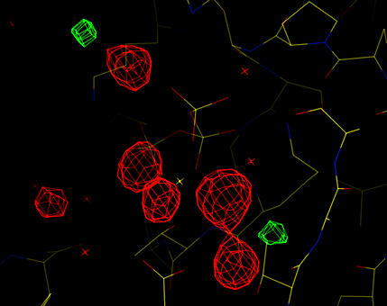
Missing waters around the magnesium ion are clear.
The model statistics are checked (as at the start of refinement) in order to check the model geometry and fit to the experimental diffraction data. In this example the CNS task file model_stats_final.inp is used to analyse the model geometry and diffraction statistics:
cns_solve < model_stats_final.inp > model_stats_final.out [2 minutes]
The output listing file (model_stats_final.list) contains a variety of information which is self-explanatory. Important things to note are:
R-values:
=================================== summary ==================================

resolution range: 500 - 1.9 A
  R-values:
    initial                                        r= 0.3787 free_r= 0.3728
    after B-factor and/or bulk solvent correction  r= 0.2535 free_r= 0.2846
R-values versus resolution:
================================= R-values ===================================


=======> R-values with |Fobs|/sigma cutoff= 0.0

 Test set (test = 1):

 #bin | resolution range | #refl |
    1   4.09  500.01        529      0.2456
    2   3.25    4.09        439      0.2512
    3   2.84    3.25        538      0.2974
    4   2.58    2.84        514      0.3214
    5   2.39    2.58        517      0.3041
    6   2.25    2.39        484      0.3032
    7   2.14    2.25        456      0.3091
    8   2.05    2.14        461      0.3490
    9   1.97    2.05        393      0.3855
   10   1.90    1.97        382      0.3589

 Working set:

 #bin | resolution range | #refl |
    1   4.09  500.01       4660      0.2028
    2   3.25    4.09       4764      0.2345
    3   2.84    3.25       4636      0.2786
    4   2.58    2.84       4532      0.2777
    5   2.39    2.58       4460      0.2809
    6   2.25    2.39       4336      0.2710
    7   2.14    2.25       4185      0.2912
    8   2.05    2.14       3993      0.3152
    9   1.97    2.05       3754      0.3212
   10   1.90    1.97       3412      0.3525
This distribution of R-values is reasonable - there is no dramatic increase in R-value (in particular free R-value) as resolution increases. If there were resolution shells with R-values significantly higher than the rest this might indicate possible problems with the data processing (ice rings for example). However, the R-values indicate the fit to the experimental data and should not be manipulated by removing data - in particular by the use of sigma cutoffs to exclude weak data.
The overall geometry:
rmsd bonds= 0.010768 with 7 bond violations > 0.05
rmsd angles=  1.30473 with 6 angle violations >  8.0
rmsd dihedrals= 21.92063 with 3 angle violations >  60.0
rmsd improper=  0.83026 with 9 angle violations >  3.0
These overall statistics are acceptable, but it is necessary to check the outlying angle and improper deviations in the detailed geometry analysis (below).
The geometry in detail:
================================= geometry ===================================

=======> bond violations

 (atom-i        |atom-j        )    dist.   equil.   delta    energy   const.

 (N    100  SD  |N    100  CE  )    1.670    1.791   -0.121    2.475  170.066
 (N    182  SD  |N    182  CE  )    1.642    1.791   -0.149    3.791  170.066
 (N    184  CG  |N    184  SD  )    1.745    1.803   -0.058    1.709  512.111
 (N    184  SD  |N    184  CE  )    1.643    1.791   -0.148    3.741  170.066
 (N    239  SD  |N    239  CE  )    1.579    1.791   -0.212    7.630  170.066
 (N    243  SD  |N    243  CE  )    1.694    1.791   -0.097    1.606  170.066
 (N    247  SD  |N    247  CE  )    1.739    1.791   -0.052    0.456  170.066

=======> angle violations

 (atom-i        |atom-j        |atom-k        )  angle    equil.     delta    energy  const.

 (N    28   N   |N    28   CA  |N    28   C   )   99.397  111.200  -11.803   10.519  247.886
 (N    58   N   |N    58   CA  |N    58   C   )  119.986  111.200    8.786    5.829  247.886
 (N    68   N   |N    68   CA  |N    68   C   )  100.627  111.200  -10.573    8.441  247.886
 (N    129  N   |N    129  CA  |N    129  C   )  102.275  111.200   -8.925    6.015  247.886
 (N    162  N   |N    162  CA  |N    162  C   )  121.455  111.800    9.655    8.831  310.948
 (N    197  N   |N    197  CA  |N    197  C   )   99.437  111.200  -11.763   10.448  247.886

=======> improper angle violations

 (atom-i        |atom-j        |atom-k        |atom-L        )    angle    equil.   delta    energy   const.   period

 (N    40   N   |N    40   CA  |N    40   CD  |N    39   C   )   -5.434    0.000    5.434    0.899  100.000   0
 (N    62   N   |N    62   CA  |N    62   CD  |N    61   C   )    4.523    0.000   -4.523    0.623  100.000   0
 (N    140  N   |N    140  CA  |N    140  CD  |N    139  C   )   -3.584    0.000    3.584    0.391  100.000   0
 (N    143  CA  |N    143  N   |N    143  C   |N    143  CB  )   38.771   35.264   -3.506    2.808  750.000   0
 (N    143  N   |N    143  CA  |N    143  CD  |N    142  C   )   -3.454    0.000    3.454    0.363  100.000   0
 (N    162  N   |N    162  CA  |N    162  CD  |N    161  C   )    5.476    0.000   -5.476    0.913  100.000   0
 (N    195  CA  |N    195  N   |N    195  C   |N    195  CB  )   38.272   35.264   -3.008    2.067  750.000   0
 (N    249  N   |N    249  CA  |N    249  CD  |N    248  C   )    5.758    0.000   -5.758    1.010  100.000   0
 (A    1    PG  |A    1    O1G |A    1    O2G |A    1    O3G )  -30.095  -35.000   -4.905    5.496  750.000   0

=======> dihedral angle violations

 (atom-i        |atom-j        |atom-k        |atom-L        )    angle    equil.   delta    energy   const.   period

 (N    182  CB  |N    182  CG  |N    182  SD  |N    182  CE  )  164.604   90.000  -74.604    9.295    5.000   2
 (A    1    O1A |A    1    PA  |A    1    O5* |A    1    C5* )  -13.796  180.000 -166.204    0.000    0.000   0
 (A    1    O5* |A    1    C5* |A    1    C4* |A    1    C3* )  -83.679  180.000  -96.321    0.000    0.000   0
Specific problems with the model geometry can be identified here. The systematically longer bond lengths for the methionine SD CE bond may seems strange. However, they are caused by the use of the experimental phases in the refinement. These phases were generated for the protein with selenomethionine. The selenium carbon bond for (SE CE) is longer and the force constant for the SD CE bond is low (allowing the bond length to deviate from the expected values).
After analysis of the geometry and fit to the experimental data electron density maps should be checked. Cross-validated, sigma-A weighted, phase combined 2Fo-Fc and difference maps are calculated:
      cns_solve < model_map_final_1fofc.inp > model_map_final_1fofc.out [30 seconds]
      cns_solve < model_map_final_2fofc.inp > model_map_final_2fofc.out [1 minute]
If you have mapman installed, you can use the command
map_to_omap *.map
to convert the CNS maps to a format which can be read into
O. In O, enter @omac to read in the current
model and map.
Analysis of the difference map shows that some ordered water molecules are still missing. These can be placed either manually or by further rounds of automated water picking.
 |
| Cross-validated, phase combined Sigma-A weighted difference map (red is +4 sigma, green is -4 sigma). Missing waters around the magnesium ion are clear. |